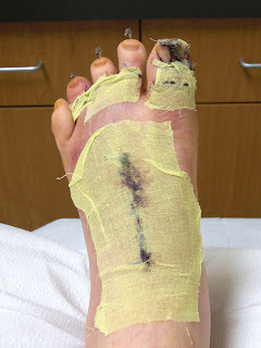
Bursitis Of The Feet Bursal Cyst
Overview
That dull misery in the shoulder, knee or elbow known as bursitis can strike anybody, from the couch potato to the highly trained athlete. Though bursitis may hurt as much as arthritis, it isn?t a joint disease. Rather, it's an acute or chronic painful inflammation of a bursa. Bursae (from the Greek word for wine-skin and related to the English word purse) are small, closed, fluid-filled sacs that protect muscles and tendons from irritation produced by contact with bones. If friction becomes too great, from overexercising, hard work, or injury, for instance-the bursae themselves may get inflamed. Though the shoulder is a common locale for bursitis, any of the bursae in the human body-there are approximately 150-can become irritated. Occupational bursitis is not uncommon and is known by old, familiar names such as "housemaid's knee," and "policeman's heel." One of the most common foot ailments, the bunion, is a form of bursitis.
Causes
Age. Bursitis is more common during middle age due to repetitive activities that put wear and tear on the body over time. Certain activities or occupations. If your job or hobby involves repetitive motion or puts pressure on bursae, you have a higher likelihood of developing bursitis. Reaching overhead, leaning elbows on arm rests, crossing your legs, laying carpet, setting tile, gardening, biking, playing baseball and ice skating are some activities that, when repeated very often, can put you at increased risk of developing bursitis. Sports in which you may get hit in the knee or fall to the knees, such as football, can also increase the risk. Some medical or health conditions. Rheumatoid arthritis, osteoarthritis, gout, thyroid disease, diabetes, alcoholism and some immunosuppressive disorders can increase the risk of bursitis. The reasons can vary, from cartilage breakdown around joints (arthritis) to crystals in the bursa that cause inflammation (gout). Wearing high heels. Posterior Achilles tendon bursitis occurs when the bursa located between the skin and the Achilles tendon (the band of tissue that attaches the calf muscle to the heel bone) becomes inflamed. High heels are often to blame for this, the stiff heel can put direct pressure on the bursa between the skin and the Achilles tendon.
Symptoms
Achiness or stiffness in the affected joint. Worse pain when you press on or move the joint. A joint that looks red and swollen (especially when the bursae in the knee or elbow are affected). A joint that feels warm to the touch, compared to the unaffected joint, which could be a sign that you have an infection in the bursa. A ?squishy? feeling when you touch the affected part. Symptoms that rapidly reappear after an injury or sharp blow to the affected area.
Diagnosis
A physical examination will be performed to determine if you have any signs of Achilles Bursitis or other ankle injury. He/she will look and feel the soft tissue and bones in your ankles to note any differences between the two of them. This will identify any abnormalities, such as swelling, bone deformities, atrophied muscles, redness and/or warmth on the skin. In many cases, the first sign that you have Achilles bursitis is swelling in the back of the foot and ankle pain.
Non Surgical Treatment
Treatments should involve decreasing swelling, relieving pain and stress on the Achilles, correcting any biomechanical dysfunction (over-pronation or flat feet), treating scar tissue, and then restoring strength and movement. If you are performing an activity that could cause further trauma to the bursa, it is recommended that you protect the area with padding and/or proper footwear to prevent further irritation or damage.
Prevention
Contact your physician if bursitis pain is disabling (when movement of the joint is largely or entirely restricted), if the pain doesn?t subside after a week of self-care, or if the joint is red and swollen. Also call your doctor if you develop a fever, which could signal infectious bursitis-a condition that especially can afflict the elbow. Except for the fever, symptoms resemble other forms of bursitis, but infectious bursitis requires immediate medical attention.
Nonsurgical Hammer Toe Repair
 Overview
Generally a hammertoe or mallet toe is caused by wearing high heels or shoes that are too small around the toe area, so it?s no surprise that it is mostly women who suffer from them. A Hammer toes has a bend in the middle joint of the toe whereas a mallet toe has a bend in the upper joint of the affected toe. The way someone walks (gait) can also lead to the formation of hammertoes and mallet toes as can overuse and injury. Sometimes a deep blister will form over the bent joint and often after some time calluses and corns will develop on the affected toe joint. People with arthritis, diabetes or neuromuscular conditions are also more likely to develop a hammer toe or mallet toe.
Causes
As described above, the main reason people develop hammertoes is improper footwear, or footwear that is too short for the toes. Shoes that do not allow our toes to lie flat are the biggest cause of hammertoes, though there are others, including genetics, injury or trauma in which the toe is jammed or broken. Diseases that affect the nerves and muscles, such as arthritis. Abnormal foot mechanics due to nerve or muscle damage, causing an imbalance of the flexor and extensor tendons of the toe. Systematic diseases such as arthritis can also lead to problems such as hammertoe. Some people are born with hammertoes, while others are more prone to developing the condition due to genetics. If you have ever broken a toe, you know there is not much that can be done for it. It is one of the only bones in the body that heals without the use of a cast. A broken toe may be splinted, however, which may help prevent a hammertoe from forming.
 Symptoms
The symptoms of a hammer toe are usually first noticed when a corn develops on the top of the toe and becomes painful, usually when wearing tight shoes. There may be a bursa under the corn or instead of a corn, depending on the pressure. Most of the symptoms are due to pressure from footwear on the toe. There may be a callus under the metatarsal head at the base of the toe. Initially a hammer toe is usually flexible, but when longstanding it becomes more rigid.
Diagnosis
A hammertoe is usually diagnosed with a physical inspection of your toe. Imaging tests, such as X-rays, may be ordered if you have had a bone, muscle, or ligament injury in your toe.
Non Surgical Treatment
There are several treatment options. These are based on how severe the problem has become. The sooner a person seeks treatment, the more options that person may have. Wear properly fitting shoes; this does not necessarily mean expensive shoes. Padding any prominent areas around the bony point of the toe may help to relieve pain. Medication that reduces inflammation can ease the pain and swelling. Sometimes a doctor will use cortisone injections to relieve acute pain. A podiatrist may also custom-make an insert to wear inside your shoe. This can reduce pain and keep the hammer toe from getting worse. Your doctor may recommend foot exercises to help restore muscle balance. Splinting the toe may help in the very early stages.
Surgical Treatment
Surgery may be the treatment of choice if conservative approaches prove unsuccessful. Usually performed as an outpatient procedure, the specific surgery will depend on the type hammertoe and extent of injury to the toe. Recovery my take several days or weeks and you may experience some redness, stiffness and swelling of the affected toe. Your physician will recommend taking it easy and to keep your foot elevated while you recover.
 Prevention
Plainly put, most toe deformities are caused by footwear. If you wish to avoid bunions or hammertoes, or works towards reversing them, please choose your footwear that has a low heel, and is wide enough in the toebox to spread you toes. Your podiatrist or therapist can help with the manipulation exercises. Bunion splints are available.
Tailor Bunions Causes
Overview
 The prominent bone at the side of the big toe rubs against the shoe and the skin becomes reddened. Often a sac of fluid called a bursa may develop in the tissue overlying the prominent bone. This swelling, consisting of inflamed soft tissues and underlying prominent bone is what we call a ?bunion?. If left untreated, they can be very painful.
Causes
Women tend to suffer from bunions more often than men, probably because of the shoes they wear. But besides shoes, standing on the feet for long periods of time can also make symptoms of a bunion worse. Along with the bump, these bunion symptoms include pain or soreness, swelling, redness around the joint, a burning sensation or sometimes numbness. The big toe may develop calluses or not be able to move as well as it once did. Sores between the toes and ingrown toenails may also occur because of a bunion.
Symptoms
Bunions may or may not cause symptoms. A frequent symptom is foot pain in the involved area when walking or wearing shoes that is relieved by resting. A bunion causes enlargement of the base of the big toe and is usually associated with positioning of the big toe toward the smaller toes. This leads to intermittent or chronic pain at the base of the big toe. Bunions that cause marked pain are often associated with swelling of the soft tissues, redness, and local tenderness. It is important to note that, in post-pubertal men and post-menopausal women, pain at the base of the big toe can be caused by gout and gouty arthritis that is similar to the pain caused by bunions.
Diagnosis
Looking at the problem area on the foot is the best way to discover a bunion. If it has the shape characteristic of a bunion, this is the first hint of a problem. The doctor may also look at the shape of your leg, ankle, and foot while you are standing, and check the range of motion of your toe and joints by asking you to move your toes in different directions A closer examination with weight-bearing X-rays helps your doctor examine the actual bone structure at the joint and see how severe the problem is. A doctor may ask about the types of shoes you wear, sports or activities (e.g., ballet) you participate in, and whether or not you have had a recent injury. This information will help determine your treatment.
Non Surgical Treatment
Currently, no strong evidence points to the best treatment for bunions. But in most cases, you can treat them at home. This includes taking medicine you can buy without a prescription to relieve toe pain. It also helps to wear shoes that do not hurt your feet. For example, avoid high heels or narrow shoes. You can wear pads to cushion the bunion, and in some cases, you can use custom-made shoe inserts (orthotics). Avoid activities that put pressure on your big toe and foot. But don't give up exercise because of toe pain. Try activities that don't put a lot of pressure on your foot, such as swimming or bicycling. Surgery to correct a bunion may be an option if other treatment does not relieve pain. There are different types of surgery for bunions. You and your doctor can decide which one is best for you. 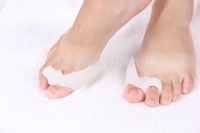
Surgical Treatment
Procedures can range from shaving off excess bone to restructuring and fusing the big toe. For mild conditions, you may simply need the connective tissues holding your big toe to be tightened so they hold the digit in the correct position. More advanced bunions will need more manipulation and involved remedies. Cuts in the bone tissue can help our specialists realign the toe. You may need to have the damaged portion of the joint removed. In severe cases, the joint may be fused to prevent it from moving out of position again. If your bunion created other foot complications, like hammertoes, our specialists may correct those during the procedure as well.
Prevention
Choosing footwear that fits correctly, especially low heeled shoes with plenty of space for the toes, is one of the main ways that bunions can be prevented. Always stand when trying on shoes to ensure they still fit comfortably when the foot expands under your body weight. Try shoes on both feet, and select the size appropriate for your larger foot. Use an extra insole if one shoe is looser than the other. Do not cramp the larger foot. People prone to flat-footedness should consider the use of arch supports, orthotic shoe inserts or special orthotic shoes to prevent or delay the development of bunions.
What Is Overpronation Of The Feet
Overview
The foot and ankle complex needs to pronate to make the muscles of the hips and legs work correctly. Many muscles that originate from the pelvis attach to both the upper and lower leg. For example, the gluteus maximus and tensor fascia latae (TFL) attach to the outside of the lower leg via the iliotibial band, while the abductors attach to the outside of the femur. When the foot pronates, the whole leg rotates inward toward the center line of the body. This inward rotation pulls the attachment of the glutes, TFL and abductors away from the origin of these muscles up on the pelvis which creates tension. Similarly, the muscles of the lower leg such as the peroneals, tibialis anterior and tibialis posterior originate on the lower leg and attach to the underside of the foot. When the foot flattens out, as it does in pronation, this pulls the insertion of these muscles away from their origin on the tibia. This action also creates tension in the muscles.
Causes
Over-pronation has different causes. Obesity, pregnancy, age or repetitive pounding on a hard surface can weaken the arch leading to over-pronation. Over-pronation is also very common with athletes, especially runners and most of them nowadays use orthotics inside their shoes. Over-pronation affects millions of people and contributes to a range of common complaints including sore, aching feet, ball of foot pain, heel Pain, achilles tendonitis, bunions, shin pain, tired, aching legs, knee pain and lower back pain. The most effective treatment solution for over-pronation is wearing an orthotic shoe insert. Orthotics correct over-pronation, thereby providing natural, lasting pain relief from many common biomechanical complaints.
Symptoms
Because pronation is a twisting of the foot, all of the muscles and tendons which run from the leg and ankle into the foot will be twisted. In over-pronation, resulting laxity of the soft tissue structures of the foot and loosened joints cause the bones of the feet shift. When this occurs, the muscles which attach to these bones must also shift, or twist, in order to attach to these bones. The strongest and most important muscles that attach to our foot bones come from our lower leg. So, as these muscles course down the leg and across the ankle, they must twist to maintain their proper attachments in the foot. Injuries due to poor biomechanics and twisting of these muscles due to over-pronation include: shin splints, Achilles Tendonitis, generalized tendonitis, fatigue, muscle aches and pains, cramps, ankle sprains, and loss of muscular efficiency (reducing walking and running speed and endurance). Foot problems due to over-pronation include: bunions, heel spurs, plantar fasciitis, fallen and painful arches, hammer toes, and calluses.
Diagnosis
So, how can you tell if you have overpronation, or abnormal motion in your feet, and what plantar fasciitis treatment will work to correct it? Look at your feet. While standing, do you clearly see the arch on the inside of your foot? If not, and if the innermost part of your sole touches the floor, then your feet are overpronated. Look at your (running/walking) shoes. If your shoes are more worn on the inside of the sole in particular, then pronation may be a problem for you. Use the wet foot test. Wet your feet and walk along a section of pavement, then look at the footprints you leave behind. A normal foot will leave a print of the heel connected to the forefoot by a strip approximately half the width of the foot on the outside of the sole. If you?re feet are pronated there may be little distinction between the rear and forefoot.
Non Surgical Treatment
Studies have shown that the most effective way to dexrease a high Q angle and lower the biomechanical stresses on the knee joint is to prevent excessive pronation with custom-maflexible orthotics. One study found that using soft corrective orthotics was more effective in reduknee pain than a traditional exercise program. A more recent study showed that Q angle asymmetries, secondary to excessive pronation affecting knee alignment, can be effectivecontrolled or corrected utilizing custom-made, flexible orthotics. Another project involving meof a running club determined that 75% of those using orthotics eliminated or greatly reduced pain in the feet, ankles, shins, knees and hips
Surgical Treatment
Calcaneal "Slide" (Sliding Calcaneal Osteotomy) A wedge is cut into the heel bone (calcaneus) and a fixation device (screws, plate) is used to hold the bone in its new position. This is an aggressive option with a prolonged period of non-weightbearing, long recovery times and many potential complications. However, it can and has provided for successful patient outcomes.
Severs Disease The Truth
Overview
Severs? disease usually presents with pain in either one or both of a sufferer?s heels. The area can be sore or tender, particularly first thing in the morning or after squeezing. Because the pain is focussed on the heel, an important part of the foot that makes contact with the ground through virtually all movement, sufferers often have to limp to alleviate their discomfort. The pain of Severs? disease is at its worst after any exertion that involves contact between a heel and the ground, particularly strenuous exercise like running or sport. The condition is caused by the wear and tear of structures in the heel, most significantly the heel bone and any attached tendons. Severs? disease is prevalent in young children who are extremely active, particularly as the heel and its attached tendons are still growing in the age group the condition most commonly affects (7-14).
Causes
Sever?s disease is most likely to occur during the growth spurt that occurs in adolescence. For girls, growth spurts usually occurs between 8 and 13 years of age. For boys, it?s typically between 10 and 15 years of age. The back of the heel hardens and becomes stronger when it finishes growing, which is why Sever?s rarely occurs in older adolescents and teenagers.
Symptoms
The pain is at the heel or around the Achilles tendon. This is felt commonly during exercise, particularly activities involving running or jumping. The back of the heel may also be tender to touch and there may be localised swelling. There may be stiffness in the calf muscles first thing in the morning and you may notice limping or a tendency to tiptoe.
Diagnosis
A physical exam of the heel will show tenderness over the back of the heel but not in the Achilles tendon or plantar fascia. There may be tightness in the calf muscle, which contributes to tension on the heel. The tendons in the heel get stretched more in patients with flat feet. There is greater impact force on the heels of athletes with a high-arched, rigid foot. The doctor may order an x-ray because x-rays can confirm how mature the growth center is and if there are other sources of heel pain, such as a stress fracture or bone cyst. However, x-rays are not necessary to diagnose Sever?s disease, and it is not possible to make the diagnosis based on the x-ray alone.
Non Surgical Treatment
Although most cases do get better on their own, recovery typically takes several weeks or months. Adolescents can continue to play sports if the activity does not cause discomfort, but staying active does prolong the recovery period without treatment. As a result, rest and avoidance of athletic activity is usually recommended, along with medication for pain and swelling.
Prevention
The best way to prevent Sever's disease is to make sure that your child wears shoes that fit properly. The heel portion of the shoe should not be too tight, and there should be good padding in the heel. It may help to put extra heel pads in your child's shoes. Some children simply get too much physical activity. For example, they may play on too many teams or practice for too long. Their heel pain is a message to slow down.
Partial Achilles Tendon Rupture Treatment Options
Overview
 A rupture of the Achilles tendon is one of the more common types of tendon ruptures. As the largest tendon in the human body, the Achilles tendon is very important to one?s balance and stability. Patients who suffer an Achilles tendon rupture experience severe pain and often require Achilles tendon surgery to repair the damage. There are also less invasive methods of Achilles tendon treatment including, icing, anti-inflammatory medications, rehabilitation, taping or bracing, sports massages and more. Achilles tendon recovery is dependent on the health of the patient, and the severity of the injury.
Causes
Achilles tendon ruptures are most likely to occur in sports requiring sudden stretching, such as sprinting and racquet sports. Achilles tendon ruptures can happen to anyone, but are most likely to occur to middle age athletes who have not been training or who have been doing relatively little training. Common sporting activities related to Achilles tendon rupture include, badminton, tennis, squash. Less common sporting activities that can lead to Achilles tendon rupture include: TKD, soccer etc. Occasionally the sufferer may have a history of having had pain in the Achilles tendon in the past and was treated with steroid injection to around the tendon by a doctor. This can lead to weakening of the tendon predisposing it to complete rupture. Certain antibiotics taken by mouth or by intravenous route can weaken the Achilles tendon predisposing it to rupture. An example would be the quinolone group of antibiotics. An common example is Ciprofloxacin (or Ciprobay).
Symptoms
Tendon strain or tendon inflammation (tendonitis) can occur from tendon injury or overuse and can lead to a rupture. Call your doctor if you have signs of minor tendon problems. Minor tenderness and possible swelling increases with activity. There is usually no specific event causing sudden pain and no obvious gap in the tendon. You can still walk or stand on your toes. Acute calf pain and swelling can indicate a tear or partial tear of the Achilles tendon where it meets the calf muscle. You may still be able to use that foot to walk, but you will need to see a specialist such as an orthopedic surgeon. Surgery is not usually done for partial tears. Sometimes special heel pads or orthotics in your shoes may help. Follow up with your doctor to check for tendonitis or strain before resuming activity, because both can increase the risk of tendon rupture. Any acute injury causing pain, swelling, and difficulty with weight-bearing activities such as standing and walking may indicate you have a tear in your Achilles tendon. Seek prompt medical attention from your doctor or emergency department. Do not delay! Early treatment results in better outcome. If you have any question or uncertainty, get it checked.
Diagnosis
Diagnosis is made by clinical history; typically people say it feels like being kicked or shot behind the ankle. Upon examination a gap may be felt just above the heel unless swelling has filled the gap and the Simmonds' test (aka Thompson test) will be positive; squeezing the calf muscles of the affected side while the patient lies prone, face down, with his feet hanging loose results in no movement (no passive plantarflexion) of the foot, while movement is expected with an intact Achilles tendon and should be observable upon manipulation of the uninvolved calf. Walking will usually be severely impaired, as the patient will be unable to step off the ground using the injured leg. The patient will also be unable to stand up on the toes of that leg, and pointing the foot downward (plantarflexion) will be impaired. Pain may be severe, and swelling is common. Sometimes an ultrasound scan may be required to clarify or confirm the diagnosis. MRI can also be used to confirm the diagnosis.
Non Surgical Treatment
Non-surgical treatment of Achilles tendon rupture is usually reserved for patients who are relatively sedentary or may be at higher risk for complications with surgical intervention (due to other associated medical problems). This involves a period of immobilization, followed by range of motion and strengthening exercises; unfortunately, it is associated with a higher risk of re-rupture of the tendon, and possibly a less optimal functional outcome. 
Surgical Treatment
Surgery is recommended to those who are young to middle-aged and active. The ruptured tendon is sewn together during surgery. This is an outpatient procedure. Afterward the leg is put into a splint cast or walking boot. Physical therapy will be recommended. In about 4 to 6 months, healing is nearly complete. However, it can take up to a year to return to sports fully.
Posterior Tibial Tendon Dysfunction Surgery Recovery
Overview
Many patients suffer from a ?collapsing arch? or ?flat foot? which can cause pain, instability and difficulty while walking. This condition is more commonly known as Posterior Tibial Tendon Dysfunction (PTTD). PTTD is a progressive flattening of the arch due to loss of function of the Posterior Tibial tendon. As the foot flattens, the tendon will stretch, become insufficient and lose its ability to function. This can have a direct effect on walking and posture, ultimately affecting the ankle, knee and hip. As the condition progresses, the joints in the hind foot may become arthritic and painful. 
Causes
Posterior tibial tendon dysfunction is the most common cause of acquired adult flatfoot. Sometimes this can be a result of specific trauma, but usually the tendon becomes injured from wear and tear over time. This is more prevalent in individuals with an inherited flat foot but excessive weight, age, and level of activity are also contributing factors.
Symptoms
The types of symptoms that may indicate Adult-Acquired Flat Foot Deformity include foot pain that worsens over time, loss of the arch, abnormal shoe wear (excessive wearing on the inner side of shoe from walking on the inner side of the foot) and an awkward appearance of the foot and ankle (when viewed from behind, heel and toes appear to go out to the side). It is important that we help individuals recognize the early symptoms of this condition, as there are many treatment options, depending upon the severity, the age of the patient, and the desired activity levels.
Diagnosis
There are four stages of adult-acquired flatfoot deformity (AAFD). The severity of the deformity determines your stage. For example, Stage I means there is a flatfoot position but without deformity. Pain and swelling from tendinitis is common in this stage. Stage II there is a change in the foot alignment. This means a deformity is starting to develop. The physician can still move the bones back into place manually (passively). Stage III adult-acquired flatfoot deformity (AAFD) tells us there is a fixed deformity. This means the ankle is stiff or rigid and doesn???t move beyond a neutral (midline) position. Stage IV is characterized by deformity in the foot and the ankle. The deformity may be flexible or fixed. The joints often show signs of degenerative joint disease (arthritis).
Non surgical Treatment
Icing and anti-inflammatory medications can reduce inflammation and physical therapy can strengthen the tibial tendon. Orthotic inserts that go inside your shoes are a common way to treat and prevent flatfoot pain. Orthotics control the position of the foot and alleviate areas of pressure. In some cases immobilization in a cast or walking boot is necessary to relieve symptoms, and in severe cases surgery may be required to repair tendon damage. 
Surgical Treatment
When conservative care fails to control symptoms and/or deformity, then surgery may be needed. The goal of surgical treatment is to obtain good alignment while keeping the foot and ankle as flexible as possible. The most common procedures used with this condition include arthrodesis (fusion), osteotomy (cutting out a wedge-shaped piece of bone), and lateral column lengthening. Lateral column lengthening involves the use of a bone graft at the calcaneocuboid joint. This procedure helps restore the medial longitudinal arch (arch along the inside of the foot). A torn tendon or spring ligament will be repaired or reconstructed. Other surgical options include tendon shortening or lengthening. Or the surgeon may move one or more tendons. This procedure is called a tendon transfer. Tendon transfer uses another tendon to help the posterior tibial tendon function more effectively. A tendon transfer is designed to change the force and angle of pull on the bones of the arch. It's not clear yet from research evidence which surgical procedure works best for this condition. A combination of surgical treatments may be needed. It may depend on your age, type and severity of deformity and symptoms, and your desired level of daily activity.